The patient

Patient right profile
Unilateral keratoconus, stable since the first visit
.
Identity : Mr T.N
First visit : 12/10/2015
Last visit : 01/08/2019
Mr. T.N, is a 24-year-old male student with no previous medical history or any known keratoconus in his family. He first came to us for a refractive surgery suitability assessment. The patient complained of a progressive decrease in visual acuity in the right eye. He also noted the perception of vertical ghosting with the right eye, which persists despite the wear of corrective spectacles for mild myopia (RE :-2.75 D, LE : -3.00D).
His refraction at the first visit (12/10th/2015) was: Right Eye (RE) 20/20 with -1.75 (-1 x 90 °) and Left Eye (LE) 20/20 with -3.25 (-0.25 x 125 °).
Clinical examination with the slit lamp revealed Vogt’s striae (fine whitish lines in the posterior stroma) and Fleischer ring (Fleischer rings are pigmented rings in the peripheral cornea, resulting from iron deposition in basal epithelial cells, in the form of hemosiderin), in the right eye.
Systematic corneal topography, performed for every refractive surgery candidate, revealed unilateral keratoconus in the right eye. The left eye topography showed only mild irregularity on the curvature map.
The patient is right-handed and sleeps on his right side with head in the pillow (« pillow hugging »).
When asked about possible rubbing habits, the patient denied eye rubbing at the first visit. His mother, however, who was with him during the consultation, claimed that she witnessed her son rubbing his eyes very often, for many years. We informed the patient that he must pay attention to the possibility of unconscious rubbing at waking up, during the day while working on the computer, at night, etc.
At the second visit, the patient confessed to becoming more aware of rubbing his eyes several times a day, especially in the mornings with his right hand, using predominantly the index pulp and knuckles. He felt an irritation on the right eye on awaking. The RE refraction was 20/20 with -2.75 (-1.00 x 100°), and the vertical ghosting persisted.
The patient’s mother took a photo of her son sleeping to demonstrate his sleeping posture, and this was presented to us.
We explained to the patient that since vigorous rubbing had preceded the drop in visual acuity and quality on the right eye, this habit may have caused the cornea to deform in his case. We strongly advised this patient to stop rubbing his eyes and to change his unhealthy sleeping position and try to sleep on the back without any ocular or orbit nocturnal compression.
Here are pictures of the patient rubbing his eyes and his sleeping posture
 Photo taken while the patient is demonstrating how he rubs his eyes (second visit).
PATIENT RUBBING HIS RIGHT EYE : Note the use of the knuckles to rub.
 This photo was taken and shared by the patient's mother. It shows the sleep position of the patient, which is invariably on the right side, with the right orbit buried in the pillow.
PATIENT SLEEP POSITION : Note the head buried deeply in the pillow (pillow hugging) and the direct contact of the pillow with the right eye (compression + heating responsible for local inflammation and irritation).
 Patient right eye profile. There is no marked "protrusion" or "distention"; rather the supero-central part of the cornea is flatter, concomitant with an inferior steepening (curvature redistribution due to the permanent corneal buckling).
  Patient left profile
 RESULTS OF E AND WHITE DISK PATCH TESTS DRAWN BY THE PATIENT AS SEEN WITH HIS RIGHT EYE). The ghosting of the E letter is inferior. The patient perceives a smeared image of the white disk, with inferior light trails and ghost images. These visual disturbances are directly caused by the asymmetrical corneal deformation.
Here is a video of the patient rubbing his right eye
Here is another video of the patient showing how he used to rub his right eye (last visit)
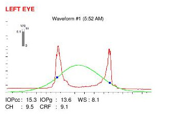
Here are the Orbscan quadmaps with SCORE ANALYZER assessment, Pentacam maps, OPDscan and Ocular Response Analyzer (ORA) results of the first visit .
Difference maps were performed at each subsequent visit. No evolution was observed between the first and last visits. The keratoconus is stable, more than 3 years after the patient definitively stopped rubbing his eyes .
At the first visit, the patient denied any excessive eye rubbing. After he received proper information, the patient acknowledged that he had come to realize his eye rubbing habit, and the preferential rubbing of the right eye. He informed us that the rubbing was so intense that it generated a « little noise », and admitted that kicking the habit was difficult to do. This stresses the importance of delivering exhaustive information to the patient and help him to realize unconscious rubbing episodes. As this cases and other demonstrate, the cessation of eye rubbing is the key factor for keratoconus stabilization.
 PENTACAM DIFFERENTIAL MAPS : RIGHT EYE (between 2nd and 3rd VISITS). The difference map demonstrates the absence of progression.
 PENTACAM DIFFERENTIAL MAPS : RIGHT EYE (between 3rd and 4th VISITS). The difference map demonstrates the absence of progression.
 ORBSCAN DIFFERENTIAL MAPS : RIGHT EYE (between 1st and 5th VISITS). The difference map demonstrates the absence of progression.
 PENTACAM DIFFERENTIAL MAPS : RIGHT EYE (between 1st and 5th VISITS). The difference map demonstrates the absence of progression.
 PENTACAM DIFFERENTIAL MAPS : RIGHT EYE (between 1st and 6th VISITS). The difference map demonstrates the absence of progression. Rather, a slight inferior flattening can be observed on the difference map (3rd column). This modification probably results from several factors; suppression of the nocturnal compression, and some degree of epithelial smoothing.
 PENTACAM DIFFERENTIAL MAPS : LEFT EYE (between 2nd and 3rd visits). The difference map shows the absence of change between the two examination time points.
 PENTACAM DIFFERENTIAL MAPS : LEFT EYE (between 3rd and 4th VISITS). The difference map shows the absence of change between the two examination time points.
 ORBSCAN DIFFERENTIAL MAPS : LEFT EYE (between 1st and 5th VISITS). The difference map shows the absence of change between the two examination time points.
It is interesting to note the concordance between unilateral rubbing of the right eye and the unilateral (right) location of the keratoconus. The patient has also been sleeping on his right side since childhood. Unilateral chronic eye irritation related to the extended compression of the right eye on the pillow induces the patient to rub his right eye during the day. At night, the right eye is exposed to local heating and contamination by irritants (allergens from laundry products, dust mites, etc.). Local heating could up regulate the activity of collagenases and related enzymes. These combined factors have facilitated the effect of repeated frictions, which eventually cause the deformation (severing of the stromal collagen lamellae) and thinning (by redistribution of the ground substance of the cornea) typically seen in keratoconus.
The deformation of the corneal dome explains the apparition of the visual disturbances (vertical ghosting of the right eye).
While he denied eye rubbing at the first visit, his mother, fortunately accompanying him, could alert him to the habit which he was not aware of. Many patients, like Mr S.M, are not conscious of their eye rubbing, or aware of its deleterious effects, and believe it to be a banal and harmless maneuver. It is important therefore, in all patients with keratoconus, to elucidate the history of eye rubbing, and then counsel the patients accordingly to change their habits.
As this case and others strongly suggest, « unilateral » keratoconus does exist: it occurs in patients who rub excessively one eye only. This is however seldom, as allergies and other factors favoring eye rubbing usually affect both eyes. In this case, the sleep position on the right side, resulting in chronic compression and heating of the cornea, along with the irritation of the eyelids and conjunctiva on the same side (promoting itch and subsequent rubbing) have synergistically contributed to the apparition of the permanent corneal thinning and warpage, which are the hallmarks of keratoconus.
In this case, like the many other cases in this website, the keratoconus ceased to evolve after the patient definitively stopped rubbing his right eye. The absence of excessive rubbing on the left eye prevented keratoconus to occur. This and other observations gathered on this website strongly suggest that keratoconus progression can be stopped and that its incidence can be suppressed, all by very simple recommendations and hygiene.


