Patient right profile
Case #32
The patient

Bilateral Asymmetric Keratoconus induced by eye rubbing
Identity : Mr J.P
First visit : 08/28/2015
Last Visit : 07/25/2017
Mr. J.P is a 34-year-old male with no previous medical history or family history of keratoconus. He has a known allergy to pollen, and complained of a progressive decrease in visual acuity in both eyes.
His refraction at the first visit (08/28th/2015) was: Right Eye (RE) 20/25 with -3 (-4 x 75 °) and Left Eye (LE) 20/25 with -4.5 (-5.5 x 10⁵ °).
Slit lamp examination revealed a keratoconus pattern with thin and irregular corneas. There were bilateral Vogt’s striae and Fleischer rings (Fleischer rings are pigmented rings in the peripheral cornea, resulting from iron deposition in basal epithelial cells, in the form of hemosiderin)
Corneal topography revealed the presence of bilateral keratoconus, more pronounced in the right eye.
When asked about his sleeping habit, the patient sleeps on his right side with the head in the pillow (« pillow hugging »). He is right-handed.
When asked about possible eye rubbing habits, the patient confessed that he rubbed his eyes especially in the mornings with his right hand, using the knuckles.
We explained to the patient that since vigorous rubbing had preceded the drop in visual acuity, this habit may have caused the cornea to deform in his case. We strongly advised this patient to stop rubbing his eyes.
Here are pictures of the patient rubbing his eyes and his profiles
 PATIENT RIGHT EYE PROFILE
 PATIENT LEFT EYE PROFILE
 PATIENT RUBBING HIS EYE WITH THE BACK OF THE HAND.
 PATIENT RUBBING HIS EYES WITH KNUCKLES
 PATIENT RUBBING HIS EYES WITH FINGERS
 PATIENT SHOWING HIS SLEEPING POSITION (RIGHT SIDE)
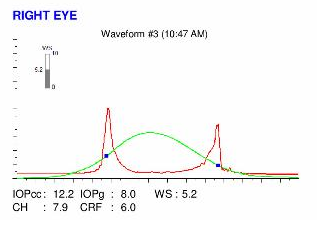
Here are the Orbscan quadmaps, Pentacam maps, OPDscan (topography and aberrometry) maps and Ocular Response Analyzer (ORA) results of the first visit .
 RIGHT EYE ORBSCAN (1st VISIT). The keratoconus is relatively centered: note the increased prolateness (negative asphericity) of the anterior (top right) and posterior (top left) corneal surfaces (marked island pattern). On the curvature map (bottom left), irregular astigmatism characterized by a marked infero-temporal steepening is obvious. The thickness map (bottom right) shows central thinning.
 LEFT EYE ORBSCAN (1st VISIT). The keratoconus is relatively centered: note the increased prolateness (negative asphericity) of the anterior (top right) and posterior (top left) corneal surfaces (marked island pattern). On the curvature map (bottom left), irregular astigmatism characterized by a marked infero-temporal steepening is obvious. The thickness map (bottom right) shows central thinning.
Difference maps were performed at each subsequent visit. No evolution was observed between the first and last visits. The keratoconus is stable, more than 2 years after the patient definitively stopped rubbing his eyes .
Rubbing your eyes is a natural reflex, a source of immediate calm, and it induces a sense of well-being. The need to rub the eyes is often a consequence of an inflammation of the ocular surface. The increase in the prevalence of allergies, the manifestations of which are accentuated or caused by the increase in atmospheric pollution, contributes to the emergence of a chronic pathology of the ocular surface. The time spent in front of the digital screen is a factor that also contributes to the emergence of a syndrome of chronic visual fatigue, which often culminates in a desire to rub the eyes. In rarer cases, as in certain conditions involving impairment of cognitive abilities (trisomy 21, autism, mental retardation), eye rubbing can become incoercible and it is in this context that the most severe forms of keratoconus are often encountered. Many of these cases eventually require a corneal transplantation.
In this case, bilateral eye rubbing with knuckles preceded the onset of keratoconus by many years. This case strongly supports our theory that eye rubbing triggers keratoconus. Cross linking is never an urgent procedure and should never be suggested immediately to a patient, as our wide experience shows that the cessation of eye rubbing is the most important first-line intervention, and a sufficient approach to stabilize corneas with keratoconus of any stage.
Other cases :
- Date 25 août 2017
- Tags Allergy, Asymmetric, Bilateral keratoconus, Eye rubbing, Fleischer ring, Male, Morning rubbing, Pillow hugging, Sleep position


