The patient

Patient right profile
Bilateral Keratoconus induced by eye rubbing
Identity : Ms I.E
First visit : 01/12/2016
Last Visit : 07/03/2018
Ms. I.E is a 29-year-old female with a history of chronic allergies in her childhood. Her brother had been recently diagnosed with keratoconus. She came to us for a second opinion, after being diagnosed with keratoconus in another hospital in Paris. She said that she had received no explanation there but was just told « you have keratoconus », and that the doctor indicated an « urgent need for cross-linking ». She was not questioned about eye rubbing habits and allergies.
Her refraction at the first visit (01/12th/2016) was : Right Eye (RE) 20/32 with -2.25 (-2.75 x 40 °) and Left Eye (LE) 20/20 with +0.25 (-2.5 x 145 °).
Clinical examination with the slit lamp revealed a suspicion of an irregular inferior corneal bulge more pronounced in the right eye. We also found bilateral Fleischer rings (Fleischer rings are pigmented rings in the peripheral cornea, resulting from iron deposition in basal epithelial cells, in the form of hemosiderin).
Corneal topography revealed the presence of bilateral keratoconus more pronounced in the right eye.
At her first visit with us, we interrogated her about her eye rubbing and sleeping habits. The patient admitted to rubbing her eyes very frequently, especially in the mornings. With regards to her sleeping habit, the patient informed us that she sleeps on her right side with her head « pushing » on the arm. She is right-handed.
We explained to the patient that since vigorous rubbing had preceded the drop in visual acuity, this habit may have caused the cornea to deform in her case. She confessed that she had rubbed her eyes very often in her childhood, as she suffered from allergic conjunctivitis. She also informed us that astigmatism was noted in her eyes when she turned 18.
We strongly advised this patient to stop eye rubbing and to change her unhealthy sleeping position to avoid chronic compression on the right eye.
Here are pictures of the patient rubbing her eyes and her profiles
 PATIENT RIGHT EYE PROFILE
 PATIENT LEFT EYE PROFILE
 PATIENT SHOWING HOW SHE RUBS HER EYES (with knuckles). This particular rubbing habit is frequently seen in patients with bilateral severe keratoconus.
 PATIENT SHOWING HER UNHEALTHY SLEEPING POSITION (head on fists)
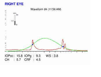
Here are the Orbscan quadmaps, Pentacam maps, OPDscan (topography and aberrometry) maps and Ocular Response Analyzer (ORA) results of the first visit.
Difference maps were performed at each subsequent visit. No evolution was observed between the first and last visits. The keratoconus is stable, more than 2 years after the patient had definitively stopped rubbing her eyes .
 PENTACAM DIFFERENTIAL MAPS : RIGHT EYE (1st to 4th VISITS). The blue color on the 3rd column suggests the presence of a spontaneous flattening! It is most presumably a fluctuation, but certainly demonstrates the stability of the keratoconus since the cessation of eye rubbing.
 PENTACAM DIFFERENTIAL MAPS : LEFT EYE (1st to 4th VISITS). The blue color on the 3rd column suggests the presence of a spontaneous flattening! It is most presumably a fluctuation, but certainly demonstrates the stability of the keratoconus since the cessation of eye rubbing.
This is a classic example of bilateral keratoconus induced by eye rubbing. Eye rubbing preceded the onset of the corneal deformation by several years. Cessation of eye rubbing resulted in the stabilization of the deformation (no keratoconus progression). The patient can next be fitted with rigid gas permeable contact lenses to restore vision.
It is important to explain to teenagers with allergies and allergic tendencies the importance of refraining from eye rubbing.


