The patient

Patient right profile
Bilateral Asymetric Keratoconus induced by eye rubbing
Identity : Mr G.Q
First visit : 06/28/2016
Last Visit : 05/22/2018
Mr. G.Q is a 41-year-old male with no previous medical history or any known keratoconus in his family. He has known allergy to dust mites, and complained of a progressive decrease in visual acuity in both eyes.
His refraction at the first visit (06/28th/2016) was : Right Eye (RE) 20/25 with -3 (-2.75 x 35 °) and Left Eye (LE) 20/25 with -3.25 (-3.50 x 130°).
Clinical examination with the slit lamp suggested an irregular inferior corneal bulge, with bilateral Vogt’s striae and Fleischer rings (Fleischer rings are pigmented rings in the peripheral cornea, resulting from iron deposition in basal epithelial cells, in the form of hemosiderin). Tarsal papillae (indicating chronic ocular allergy) were observed on the inner surface of the eyelids.
Corneal topography revealed the presence of bilateral keratoconus more pronounced in the left eye.
When asked about the possibility of frequent eye rubbing, the patient admitted to rubbing his eyes frequently with his knuckles. He sleeps on his left side.
We explained to the patient that since vigorous rubbing had preceded the drop in visual acuity, this habit may have caused the cornea to deform in his case. We strongly advised him to stop rubbing his eyes. We also recommended that he change his sleeping position, as extended compression of the eye at night time may cause local irritation and increase contamination by dust mites and irritants, which trigger more pruritus and subsequent eye rubbing.
Here are pictures of the patient rubbing his eyes and his profiles
 PATIENT RIGHT EYE PROFILE
 LEFT EYE PROFILE. Note the injection and congestion of the conjunctival vessels in the temporal bulbar conjunctiva. This reflects a chronic inflammation of the ocular surface. This inflammation is probably related to his unhealthy sleeping position.
 PATIENT DEMONSTRATING HOW HE RUBS HIS EYES (USING HIS FIST AND PALM). The patient declares that he compresses his left eye intensely, with circular and grinding movements.
 PATIENT SHOWING HIS SLEEPING POSITION. The prolonged compression of the left eye, the contamination of the ocular surface with dust mites from the linen and other irritants causes chronic irritation in the eye. The patient admits to rubbing his eyes vigorously in the morning, especially the left.
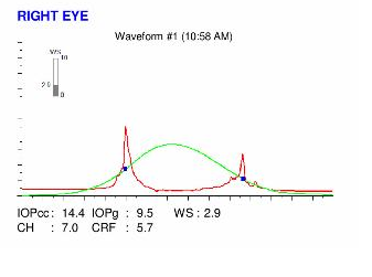
Here are the Orbscan quadmaps, Pentacam maps, OPDscan (topography and aberrometry) maps, Ocular Response Analyzer (ORA) AND OCT VISANTE results of the first visit .
 RIGHT EYE OPDscan (1st VISIT). The wavefront analysis reveals an increase in higher order aberrations (trefoil, coma, negative spherical aberration) which is the direct consequence of the loss of regularity of the corneal surface. These aberrations explain the loss of visual quality, which cannot be improved with spectacles.
Difference maps were performed at each subsequent visit. No evolution was observed between the first and last visits. The keratoconus is stable, more than 9 months after the patient definitively stopped rubbing his eyes .
 PENTACAM DIFFERENTIAL MAPS : RIGHT EYE (1st and 2nd VISITS). The subtraction map reveals the absence of progression between the 1st and second visits.
 PENTACAM DIFFERENTIAL MAPS : RIGHT EYE (2nd and 3rd visits ). The difference map demonstrates objectively the absence of keratoconus progression (3rd column).
 PENTACAM DIFFERENTIAL MAPS : LEFT EYE (1st and 2nd VISITS). The keratoconus in the left eye is stable between the 1st and 2nd visits.
 PENTACAM DIFFERENTIAL MAPS : LEFT EYE (2nd and 3rd VISITS). As for the left eye, the subtraction map reveals the absence of keratoconus progression between the 2nd and 3rd visits.
This is a case of advanced keratoconus caused by chronic vigorous eye rubbing. The eye rubbing preceded the appearance of keratoconus by several years. As in many other cases, there is a correlation between the sleeping position (side) and the laterality of the more advanced keratoconus. Fortunately for this patient, the eye rubbing habit was identified and the disease progression arrested once the patient stopped rubbing his eyes.
The origin of keratoconus remains relatively unknown. However, many observations including this case report and the other cases in this website accredit eye rubbing to be the number one causal suspect. The deformation induced by rubbing is possibly aggravated by a co-existent chronic nocturnal compression (pressure of the pillow on the eye), which itself is a source of chronic irritation and contamination, resulting in pruritus and subsequent rubbing in response to the ocular discomfort.


