The patient

Patient right profile
Unilateral Asymmetric Keratoconus induced by eye rubbing
Identity : Mr C.M
First visit : 01/17/2017
Last Visit : 05/15/2018
Mr. C.M is a 25-year-old male with no previous medical history or family history of keratoconus. He had consulted ophthalmologists from another institution and was advised to have corneal collagen cross-linking. He came to us for a second opinion. He complained of a progressive bilateral decrease in visual acuity, more pronounced in the right eye than the left.
His refraction at the first visit (01/17th/2017) was : Right Eye (RE) 20/60 with -0.25 (-4.5 x 30 °) and Left Eye (LE) 20/25 with -1.25 (-1.25 x 160 °).
Clinical examination with the slit lamp revealed an irregular inferior corneal bulge in the right eye .We also found a Fleischer ring (Fleischer rings are pigmented rings in the peripheral cornea, resulting from iron deposition in basal epithelial cells, in the form of hemosiderin) in the right eye.
When asked about the possibility of frequent eye rubbing at the first visit, he admitted that he rubbed his eyes but only occasionally, especially the right eye with his right hand (he is right-handed). However, his mother, who was present at the consultation and examination confirmed that she often witnessed her son rubbing his eyes vigorously. With regards to his sleeping posture, she said that he slept on his stomach with his head in the pillow (pillow hugging) and had a tendency to bury his eyes and orbits deeply in the pillow.
Corneal topography revealed the presence of unilateral keratoconus in the right eye.
We explained to the patient that since vigorous rubbing had preceded the drop in visual acuity, this habit may have caused the cornea to deform. We strongly advised this patient to stop the eye rubbing and to change his unhealthy sleeping position.
Here are pictures of the patient rubbing his eyes and his profiles
 PATIENT RIGHT EYE PROFILE
 PATIENT LEFT EYE PROFILE
 PATIENT RUBBING HIS EYES. This rubbing habit can be detrimental especially when the pressure exerted on the globe is particularly elevated.
 PATIENT DEMONSTRATING HIS SLEEPING POSITION. He presses the right side of his head and eye against his right hand and pillow.
 THE MOTHER DEMONSTRATING HOW HER SON RUBS HIS EYES (AND STRESSING THAT HE RUBS MUCH MORE OFTEN THAN HE THINKS).
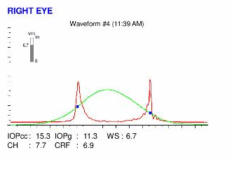
 Here are the Orbscans quadmaps with SCORE ANALYSER results, Pentacams, OPDscan (topography and aberrometry) maps, Ocular Resonse Analyzer (ORA) and OCT results of the first visit .
Difference maps were performed at each subsequent visit. At the second visit, the patient acknowledged that he had minimized the amount of rubbing, particularly in the right eye, as he understood the direct causal effect of this habit on corneal deformation in his case. No progression of keratoconus was observed between the first and last visits. The keratoconus remained stable more than 8 months after the patient had definitively stopped rubbing his eyes .
 PENTACAM DIFFERENTIAL MAPS : RIGHT EYE. The difference maps demonstrates the absence of progression.
 PENTACAM DIFFERENTIAL MAPS : RIGHT EYE. This subsequent difference map demonstrates the stability of the deformation in the right eye, consecutive to the cessation of eye rubbing).
In this case we find many triggers for eye rubbing like extended computer work and an unhealthy sleeping position. The asymmetry of keratoconus development may be related to the sleeping position (right sided) and the habit of preferentially rubbing the right eye. This patient is right-handed, and we believe that the right-hand dominance here may have a role as well in the development of asymmetry in the keratoconus disease process. When a person rubs his eyes with the dominant hand, the chances of ocular contamination with germs or allergens are higher since this hand is « more used » (as dominant) and therefore more prone to touching things, and being contaminated. Thus more itch and ocular surface irritation results. The force applied during rubbing is also probably higher than with the non-dominant hand, as the dominant hand is « stronger ».
The presence of a relative during the visit can help to confirm or identify the presence of a chronic eye rubbing habit.
This case is very informative and demonstrative of the causal effects of eye rubbing in the pathogenesis of keratoconus. Cross-linking is unnecessary in this case, as stabilisation of the corneal deformation was achieved with the simple act of cessation of eye rubbing.
As demonstrated again in this clinical example, the cessation of eye rubbing and patient education are the best tools in the prevention of the genesis and/or evolution of keratoconus.


