Patient right profile
Case #51
The patient

Bilateral Keratoconus induced by eye rubbing
Identity : Mr M.T
First visit : 06/28/2016
Last Visit : 03/20/2018
Mr. M.T is a 26-year-old male trader with allergy to pollen. He has no known history of keratoconus in his family. He complained of a progressive decrease in visual acuity greater in the right eye than the left eye.
His refraction at the first visit at the Rothschild foundation (on 06/28th/2016) was : Right Eye (RE) 20/25 with -1.75 (-4.75 x 35 °) and Left Eye (LE) 20/60 with -2.5 (-7.25 x 155 °).
Clinical examination with the slit lamp revealed bilateral Vogt’s striae, Fleischer ring and tarsal papillae. Fleischer rings are pigmented rings in the peripheral cornea, resulting from iron deposition in basal epithelial cells, in the form of hemosiderin. Vogt’s striae are thin vertical streaks located in the posterior corneal stroma (at the level of the Descemet membrane).
Corneal topography performed at our institution showed the presence of a bilateral keratoconus, more pronounced in left eye.
We investigated the risks factors for eye rubbing at the first visit. The patient disclosed known allergy to dust mites and pollen. He admitted to rubbing his eyes intensively during his teenage years especially during his many allergic crises, and in the mornings when he awakes.
With regards to his sleeping habit, the patient informed us that he is left-handed and sleeps on his left side.
At the subsequent visits, he recalled rub his eyes sometimes heavily, before his teenage years, during childhood, due again to some allergic episodes. He also became conscious of rubbing his eyes at work, where the environment was dusty, and he had since stopped after his first visit.
We explained to the patient that since vigorous rubbing had preceded the drop in visual acuity, this habit may have caused the cornea to deform, leading to the classic clinical presentation of keratoconus. The patient agreed that this pathway was logical and could well explain his keratoconus history.
We strongly advised this patient to stop the eye rubbing, to change his unhealthy sleep position and to seek the opinion of a doctor specialising in allergies .
Here are pictures of patient rubbing his eyes and profiles
 Patient right eye profile.
 Patient left eye profile
 Patient rubbing his eyes with knuckles
 Patient showing his sleep position (on sides)
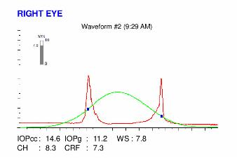
Here are the Orbscan quadmaps, Pentacam exams, OPD scans and Ocular Resonse Analyzer (ORA) results of the first visit .
Difference maps were performed at each subsequent visit. No evolution was observed between the first and last visits. The keratoconus is still stable, more than 15 months after the patient had definitively stopped rubbing his eyes.
Chronic eye rubbing can reduce the biomechanical resistance of the collagen fibers of the corneal dome and lead to the deformation of the latter. This biomechanical mechanism is more likely to account for the disparity between right and left eye involvement (patients often rub one eye more than the other), and the focal nature of keratoconus, which has recently been evidenced.
In our experience, the cessation of eye rubbing is the most important parameter in the control of the progression of corneal deformation. In our opinion, keratoconus is not an inherited disease, but the consequence of repeated mechanical trauma. Logically, the cessation of trauma leads to the eradication of the cause of the deformation and thus like many cases on this site, the cessation of eye rubbing arrests the evolution of keratoconus. This website provides many other encouraging examples of this.
Autres cas :
- Date 8 octobre 2017
- Tags Allergy, Bilateral keratoconus, Childhood rubbing, Dry eyes, Enjoyed eye rubbing, Eye rubbing, Fleischer ring, Inferior keratoconus, Knuckles rubbing, Lens removal rubbing, Male, Morning rubbing, Sleep position, Work rubbing


