Patient right profile
Case #56
The patient

Bilateral Asymmetric Keratoconus induced by eye rubbing
Identity : Mr B.E
First visit : 05/30/2017
Last Visit : 10/17/2017
Mr. B.E is a 25-year-old male with no family history of keratoconus. He gave a birth history of prematurity but with no consequent medical or surgical issues. He consulted us because of a progressive decrease in visual acuity greater in the left eye than the right, and frequent redness of the left eye.
His refraction at the first visit at the Rothschild Foundation (on 05/30th/2017) was : Right Eye (RE) 20/200 with -2.75 (-8.25 x 45 °) and Left Eye (LE) 20/60 with -2.5 (-6.75 x 135 °).
Clinical examination with the slit lamp revealed bilateral Vogt’s striae and Fleischer rings. (Fleischer rings are pigmented rings in the peripheral cornea, resulting from iron deposition in basal epithelial cells, in the form of hemosiderin. Vogt’s striae are thin vertical streaks located in the posterior corneal stroma (at the level of the Descemet membrane)). We also found superficial punctate keratopathy in the inferior cornea of the left eye.
Corneal topography performed at our institution showed the presence of bilateral keratoconus more pronounced in the left eye.
When asked about the possibility of frequent eye rubbing, the patient admitted to rubbing his eyes while watching television, as it gives him a sense of well-being. He has no known allergis. He sleeps on his left side with his head on the arm. He often awakes with bilateral eye redness, left eye more than the right.
We explained to the patient that since vigorous rubbing had preceded the drop in visual acuity, this habit may have caused the cornea to deform, leading to the classic clinical presentation of keratoconus in his case.
We strongly advised him to stop rubbing his eyes and to change his unhealthy sleeping position. We also prescribed him an eye shield for the left eye for use during sleep. We treated his dry eyes with artificial tears and vitamin A ointment at night.
Here are pictures of the patient rubbing his eyes and his profiles
 Patient right profile
 Patient left profile. Note the position of the eyelashes (pointing downwards due to chronic eye rubbing)
 Patient sleeping position (on left side)
 Note the conjunctival hyperaemia in the left eye
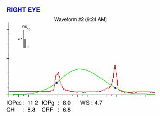
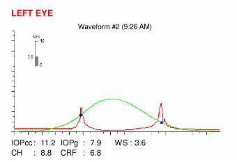
Here are the Orbscan quadmaps, Pentacam exams, OPD scans and Ocular Response Analyzer (ORA) results of the first visit .
Difference maps were performed at each subsequent visit. No evolution was observed between the first and last visits. The keratoconus is stable, more than 5 months after the patient definitively stopped rubbing his eyes .
It is of paramount importance to determine the triggers for eye rubbing in every case. In this case it was severe dry eye inducing ocular surface changes with inferior punctate keratopathy. Treatment of the ocular surface with copious lubricants addresses the ocular discomfort and the need to rub the eyes. This, together with sensitization of the patient to the deleterious effects of eye rubbing, and the actual act of cessation of rubbing was enough to arrest the progression of keratoconus in his case.
This case also reveals the relevance of the sleeping posture in the genesis and evolution of keratoconus.
Indeed, some sleep positions are more detrimental to the cornea(s) than others. The association between such sleeping positions with keratoconus (on the eye that is compressed) is striking and often underestimated. An unhealthy sleeping posture can itself cause a direct deformation of the cornea (by pressure, friction or palpebral malposition during night time). It could also cause eye irritation, inducing itch and hence the need for rubbing in the mornings. In the long term, weakening of corneal biomechanics leads to corneal thinning and curvature changes, culminating in the picture of keratoconus.
At the time of diagnosis, every patient with known or suspected keratoconus should be questioned about his sleeping habits and postures. It is important to sensitize the patient to the dangers of an unhealthy sleeping position, to modify his habits, so as to prevent through the cessation of eye rubbing, the evolution of keratoconus.
Other cases :
- Date 5 novembre 2017
- Tags Asymmetric, Bilateral keratoconus, Dry eyes, Enjoyed eye rubbing, Eye rubbing, Eye shield, Fleischer ring, Male, Sleep position, Vogt's striae


