Patient right profile
Case #77
The patient

Bilateral Asymmetric Keratoconus induced by eye rubbing
Identity : Mr M.A
First visit : 05/19/2017
Last Visit : 12/08/2017
Mr. M.A is a 25-year-old male with no known family history of keratoconus. He complained of a progressive decrease in visual acuity greater in the right eye than the left, which started at the age of 19. The patient has allergies to cat’s hair and pollen, and he rubbed his eyes very frequently during childhood because of this allergy.
He was corrected for mild hyperopia with spectacles in his earlier years and does not remember having been highly astigmatic until later. His vision started to degrade at the age of 19, but he was only diagnosed with keratoconus in February 2017 at another center. The doctors there noted that the keratoconus was progressing and so offered him corneal collagen cross-linking to stabilize the cornea. As he did not want to have the procedure performed, he consulted us for a second opinion. He did not receive specific information about eye rubbing from the doctors who diagnosed the keratoconus.
His refraction at the first visit at the Rothschild Foundation (on 05/19th/2017) was : Right Eye (RE) 20/60 with +4.25 (-5.5 x 75 °) and Left Eye (LE) 20/32 with +2 (-2.0 x 85 °).
Clinical examination with the slit lamp suggested a thin and irregular right cornea with Fleischer ring. Fleischer rings are pigmented rings in the peripheral cornea, resulting from iron deposition in basal epithelial cells, in the form of hemosiderin.
Corneal topography performed at our institution showed the presence of bilateral keratoconus, more pronounced in the right eye.
At the first visit, when asked about the possibility of frequent eye rubbing, the patient admitted to rubbing his eyes since the age of 10, because of seasonal allergic conjunctivitis (pollen allergy). When asked in more detail about his eye rubbing habits, he declared that when he awoke in the mornings or when working in front of the computer, he would rub his eyes frequently because he enjoyed it. Rubbing his right eye provided him with a sense of relief and calm.
He is right handed, and always sleeps on the right side, with the head buried in the pillow (pillow hugging).
He has a pet cat, despite being allergic to cat hair.
We strongly advised him to stop rubbing his eyes and to change his unhealthy sleeping position. We also explained to the patient that since vigorous rubbing had preceded the drop in visual acuity, this habit may have caused the cornea to deform, leading to the classic clinical presentation of keratoconus in his case.
At the subsequent visits, he admitted to not being able to successful curb his urge to rub his eyes. He was also still sleeping on his right eye and pillow hugging. His eyes were constantly itchy due to the allergy to his cat’s fur, and he would often have to rub his eyes vigorously after a shower or when in front of the computer. He also admitted to not being able to stop rubbing his eyes because he enjoyed it, and that the eye drops prescribed did not lessen the itch. He told us that he felt it was less important for him to stop rubbing his right eye because the vision of this eye was already poor.
For visual rehabilitation, we proposed fitting him with a scleral contact lens.
Here are pictures of the patient rubbing his eyes and his profiles
 PATIENT RIGHT PROFILE. You can note the change of corneal curvature occurring in the inferior aspect of the cornea.
 PATIENT LEFT PROFILE. Same as right eye, the steepened inferior aspect of the cornea is perceptible but less prominent than in the right eye..
 PATIENT RUBBING HIS RIGHT EYE WITH HIS FINGER PULP (WITH HIGH PRESSURE). The patient admits to rubbing his eyes vigorously to relieve the itch from the allergy to his cat's fur, and also because of the pleasurable sensations derived from eye rubbing.
  PATIENT DEMONSTRATING HIS SLEEPING POSITION (ON THE RIGHT SIDE). This sleeping position is likely to be playing a key role in keratoconus genesis in his case.. Chronic night-time ocular compression and irritation results in chronic inflammation and itch, which incites the eye rubbing process.
Patient showing how he rubs his right eye with the pulp of his fingers (inducing high pressure on the eyeball and cornea)
Here is a video of the patient demonstrating how he rubs his eyes with his glasses on (resulting in the « offset glasses » sign)
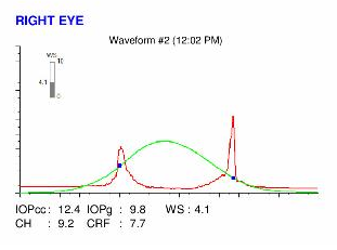
Here are the Orbscan quadmaps, Pentacam maps, OPD scans and Ocular Response Analyzer (ORA) results of the first visit .
 RIGHT EYE OPDscan examination (corneal topography and total eye wavefront aberrometry). There is an important increase in higher order aberrations (coma and trefoil), caused by the corneal distortion. The corneal inferior steepening induces a marked myopic shift in the refraction of the lower half of the pupil. The reduction of the optical quality of this eye explains why the patient cannot experience satisfactory vision with spectacle correction.
 LEFT EYE OPDScan. There is an important increase in higher order aberrations, caused by the corneal distortion. The corneal inferior steepening results in an increase of the optical power of the eye (myopic shift in the lower half of the pupil). There is a marked increase of local myopia. This asymmetry, which can be also described as an increase in aberrations such as coma and trefoil is responsible for the reduction of the quality of vision.
 Difference maps were performed at each subsequent visit. At the second visit, which was planned a month later but occurred 3 months later as the patient forgot his appointment, the patient confessed that he was rubbing his right eye even more than he had previously admitted to, at times for 10 seconds or more continuously, and about 10 times an hour. At the third visit, he admitted that he could not stop rubbing his eyes, especially the right eye, even if he fully knew that this would result in a progression of the deformation. As his right eye vision was so degraded, he declared that the cessation of eye rubbing would, in his opinion, result in no significant change in his life, but instead deprive him of a pleasurable habit…
With this refusal to stop the eye rubbing habit, we offered the patient corneal collagen cross-linking. He refused, and stated clearly his desire not to have any surgical intervention in either eye.
 PENTACAM DIFFERENTIAL MAPS : LEFT EYE. (between first and 3rd visits). This difference map now clearly shows the presence of keratoconus progression. This progression is less obvious than in the right eye, as the patient tries to avoid eye rubbing the left eye as much as he can. However, he admits that he may continue to rub his left eye as well, especially in the mornings.
This interesting case illustrates that the progression of keratoconus is directly related to the persistence of eye rubbing. Unlike the other cases in this website where the patients are able to refrain from rubbing their eyes, this patient unfortunately, is unable to stop. The responsible allergen (his cat’s hairs) is persistently in the environment, triggering an allergic cascade and the need to rub his eyes constantly to relieve the itch. Long hours spent in front of the computer screen, which causes visual fatigue associated with dry eye (reduced blinking), and an unhealthy sleeping position are also factors. The ocular discomfort in such instances is often relieved (transiently) by eye rubbing, which as described by patients, can be pleasurable and relaxing in such circumstances. Currently, only one other case in this website has shown progression of keratoconus, again as a consequence of reduced compliance and persistent eye rubbing. (Down syndrome)
All the other cases presented in this site, which we have established to have non-progressive or stable keratoconus, have only one common point: the definitive cessation of eye rubbing. This necessarily involves the removal of all the factors that trigger eye rubbing including eradication of the responsible allergens, changing the sleeping posture and / or improving work habits. As long as one of these factors persists, and the patient is not made aware of his eye rubbing habit, the progression of keratoconus is inevitable.
Our goal is not to victim-blame those who are progressing, but to inform and educate the patients to better understand the circumstances surrounding the genesis and evolution of their keratoconus, in order to help them to stop rubbing their eyes, as as to stabilize their illness.
The corneal dome can be likened to a shell whose equilibrium geometry depends on the difference between the intraocular pressure (exerted on its posterior surface) and the atmospheric pressure. Beyond a certain threshold, the mechanical stresses (compression, shear) conveyed by eye rubbing results in a biomechanical embrittlement, due to the rupture of the harmonious arrangement of collagen fibers, which causes an irreversible deformation of the cornea (This mechanism is analogous to the « buckling » in resistance of materials). If a constant pressure is exerted on the cornea, the deformation can only get worse.
Stabilization of keratoconus is therefore most challenging in these cases, and this necessarily involves the definitive cessation of eye rubbing.
Other cases :
- Date 15 février 2018
- Tags Allergy, Asymmetric, Bilateral keratoconus, Computer screen, Enjoyed eye rubbing, Eye rubbing, Eye shield, Inferior keratoconus, Male, Nails rubbing, Post shower rubbing, Progression, Sleep position, Tap water, Work rubbing


