Patient right profile
Case #80
The patient

Bilateral Asymmetric Keratoconus induced by eye rubbing
Identity : Mr H.W
First visit : 09/19/2017
Last Visit : 03/06/2018
Mr. H.W is a 22-year-old male with no previous medical history. He complained of a progressive decrease in visual acuity greater in the right eye than the left. The diagnosis of keratoconus was made in another institution, and he was advised to have corneal collagen cross-linking performed. He was not questioned on his eye rubbing habits by the ophthalmologists there. With regards to family history, the patient has an aunt who had corneal transplantation performed for keratoconus.
His refraction at the first visit at the Rothschild Foundation (on 09/19th/2017) was : Right Eye (RE) 20/25 with+0.25 (-2.5 x 70 °) and Left Eye (LE) 20/20 with -0.5 (-0.75 x 115 °).
Clinical examination with the slit lamp suggested bilateral thin and irregular corneas with a Fleischer ring in the right eye (Fleischer rings are pigmented rings in the peripheral cornea, resulting from iron deposition in basal epithelial cells, in the form of hemosiderin) and Vogt’s Striae in both eyes. Both eyes were dry with tear film break up time < 8 secs.
Corneal topography performed at our institution showed the presence of bilateral keratoconus, more pronounced in the right eye.
At the first visit, when asked about the possibility of frequent eye rubbing, the patient admitted to rubbing his eyes when he awoke in the mornings and when working in front of the computer. He found eye rubbing enjoyable.
He is left handed but rubs his right eye with his right hand. The patient sleeps on his right side, with the head buried in the pillow (pillow hugging).
We explained to the patient that since vigorous rubbing had preceded the drop in visual acuity, this habit may have caused the cornea to deform, leading to the classic clinical presentation of keratoconus in his case.
We thus strongly advised him to stop rubbing his eyes and to change his unhealthy sleeping position.
At the subsequent visits, he was accompanied by his parents who described in detail how their son would rub his eyes frequently and vigorously with the knuckles, rubbing especially his right eye with his right hand. They also confirmed his predominant sleeping position on the right side (see picture below).
After his first and subsequent consultations, the patient became conscious of his eye rubbing habits and therefore made an effort to stop. He was also prescribed an eye shield to be worn at night, and lubricant eye drops to address his dry eye problem, and to curb his desire to rub his eyes.
Here are pictures of the patient rubbing his eyes and his profiles
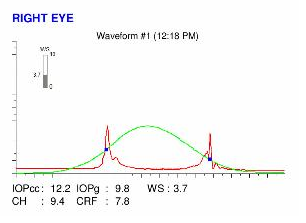
Here are the Orbscan quadmaps, Pentacam maps, OPD scans, Ocular Response Analyzer (ORA) and Corneal OCT (cross sectional and epithelial) maps of the first visit .
Difference maps were performed at each subsequent visit. No evolution has been observed between the first and last visits. The keratoconus is stable, more than 6 months after the patient definitively stopped rubbing his eyes .
This case demonstrates that eye rubbing is often triggered by long hours spent in front of the computer, which causes visual fatigue associated with dry eye (Computer Vision Syndrome). Eye rubbing is also associated with an unhealthy sleeping position, where night-time ocular compression from sleeping on the eye or orbit induces local irritation and the desire to rub upon awakening. These sensations of fatigue and irritation are often relieved (transiently) by eye rubbing, which as described by patients, can be pleasurable and relaxing in such circumstances.
The corneal dome can be likened to a shell whose equilibrium geometry depends on the difference between the intraocular pressure (exerted on its posterior surface) and the atmospheric pressure. Beyond a certain threshold, the mechanical stresses (compression, shear) conveyed by eye rubbing results in a biomechanical embrittlement, due to the rupture of the harmonious arrangement of collagen fibers, which causes an irreversible deformation of the cornea (This mechanism is analogous to the « buckling » in resistance of materials).
The repeated and sustained frictions from eye rubbing over the long-term are responsible for a permanent warpage of the cornea, culminating in the condition called « keratoconus ». Rubbing with the knuckles is particularly detrimental to the corneas, because the knuckles are the hardest part of the hands.
Since the cessation of rubbing, there has been no aggravation of the keratoconus in this patient
As demonstrated again in this clinical example, the cessation of eye rubbing and patient education are the best tools in the prevention of the genesis and/or evolution of keratoconus.
Other cases :
- Date 10 mars 2018
- Tags Asymmetric, Bilateral keratoconus, Computer screen, Enjoyed eye rubbing, Eye rubbing, Inferior keratoconus, Knuckles rubbing, Male, Morning rubbing, Offset glasses, Pillow hugging, Post shower rubbing, Sleep position, Stabilization, Witness, Work rubbing


