Patient right profile
Case #62
The patient

Unilateral Asymmetric Keratoconus induced by eye rubbing
Identity : Mr H.G
First visit : 09/12/2017
Last Visit : 10/31/2017
Mr. H.G is a 26-year-old male computer engineer with asthma and allergies commencing during childhood. There is no known family history of keratoconus. He complained of a progressive decrease in visual acuity greater in the left eye than the right for 3 years.
His refraction at the first visit at the Rothschild Foundation (on 09/12th/2017) was : Right Eye (RE) 20/25 with+0.25 (-0.5 x 90 °) and Left Eye (LE) 20/32 with +1.5 (-2.75 x 125 °).
Clinical examination with the slit lamp suggested a thin and irregular left cornea with Fleischer ring and Vogt’s striae. Fleischer rings are pigmented rings in the peripheral cornea, resulting from iron deposition in basal epithelial cells, in the form of hemosiderin. Vogt’s striae are thin vertical streaks located in the posterior corneal stroma (at the level of the Descemet membrane). These folds disappear with external gentle pressure on the globe.
Corneal topography performed at our institution showed the presence of keratoconus in the left eye and forme fruste keratoconus (FFKC) in the right eye.
At the first visit, when asked about the possibility of frequent eye rubbing, the patient admitted to rubbing his eyes frequently since childhood. He would rub with his knuckles, especially in front of the computer at night, because he found it enjoyable.
At the second visit, he described to us his frequent allergic episodes during childhood when he would rub his eye vigorously, especially in the mornings upon awakening.
While at the computer, with his right hand on the mouse, he would preferentially rub his left eye with his left hand. He sleeps on his left side, with the head buried in the pillow (pillow hugging).
We explained to the patient that since vigorous rubbing had preceded the drop in visual acuity, this habit may have caused the cornea to deform, leading to the classic clinical presentation of keratoconus in his case.
We strongly advised this patient to stop rubbing his eyes and to change his unhealthy sleeping position.
Here are pictures of the patient rubbing his eyes and his profiles
 PATIENT RIGHT PROFILE
 PATIENT LEFT PROFILE
 PATIENT RUBBING HIS EYES WITH HIS KNUCKLES
 PATIENT DEMONSTRATING HIS SLEEPING POSITION (ON HIS LEFT SIDE)
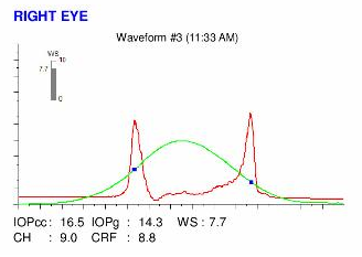
Here are the Orbscan quadmaps, Pentacams, OPD scans, Ocular Response Analyzer (ORA) and OCT VISANTE with epithelial map results of the first visit.
 LEFT EYE PENTACAM (1st VISIT). The axial curvature map (top left) shows the presence of marked vertical asymmetry. Central thinning is obvious on the thickness map (bottom left), Hyperprolateness and asymmetry is also perceptible on the anterior elevation (top right) and posterior elevation (bottom right) maps.
 LEFT EYE OPDscan map examination. The corneal deformation causes the ocular wavefront to be distorted (coma, negative spherical aberration, trefoil). These aberrations cannot be corrected by spectacles. This causes the vision to remain affected even with the "best spectacle correction". The perception of "vertical tails" around bright lights in common at night.
Difference maps were performed at each subsequent visit. No evolution has been observed between the first and last visits. The keratoconus is stable, more than 1 month after the patient definitively stopped rubbing his eyes .
This case is typical of focal corneal deformation induced by chronic eye rubbing that commenced in childhood. Very often, like many other cases on this site, the eye rubbing begins in childhood and is related to allergy. During childhood, the cornea is more deformable and the friction applied is often more intense. Patients will usually notice a decline in vision about 3 years after the onset of intense rubbing.
In this case we find many triggers for eye rubbing like extended hours of work on the computer and an unhealthy sleeping position. The unilateral or asymmetric nature of keratoconus development may be related to the sleeping position (left sided) and the habit of preferentially rubbing the left eye. .
This case is very informative and demonstrative of the causal effects of eye rubbing on the pathogenesis of keratoconus. Performing cross-linking right after the first visit seems unnecessary in this case, as the early stabilization of the corneal deformation was achieved with the simple act of cessation of eye rubbing. Longer follow-up is of course needed, and the result of further examinations will be reported here.
In our experience, the cessation of eye rubbing is the most important parameter in the control of the progression of corneal deformation. Also, allergy must be detected and treated aggressively.
The key in the fight against keratoconus is prevention by educating patients, including parents and their children about the dangers of eye rubbing.
Autres cas :
- Date 9 décembre 2017
- Tags Allergy, Asymmetric, Childhood rubbing, Computer screen, Enjoyed eye rubbing, Eye rubbing, Forme frustre keratoconus (FFKC), Knuckles rubbing, Male, Morning rubbing, Night shift, Sleep position, Unilateral keratoconus, Vogt's striae, Work rubbing


